
Fig 5. Effect of HK I or II over-expression on mitochondrial associated HK, organelle injury, caspase 3 activation and cell survival after stress.
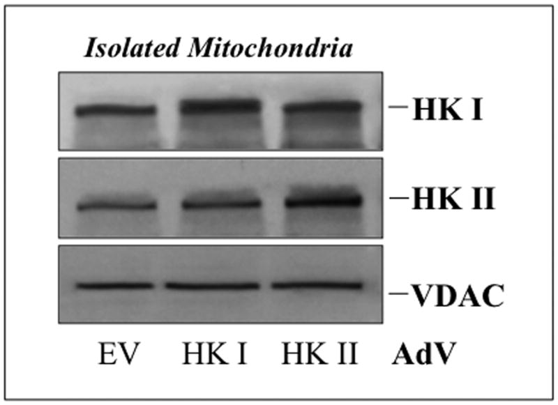
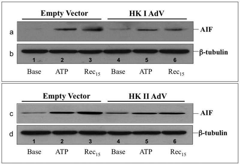
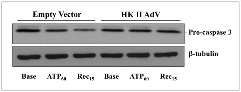
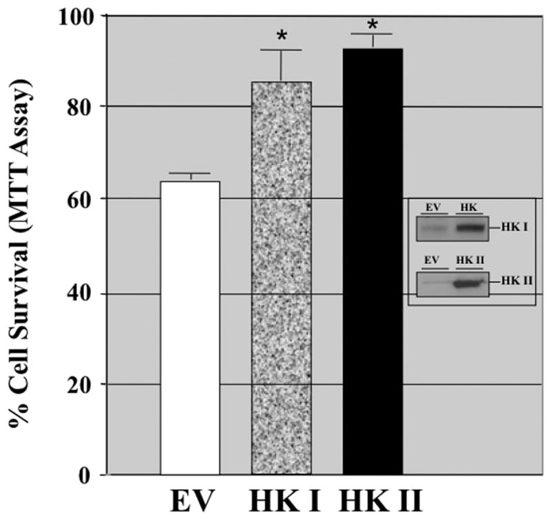
(A) HK I and HK II content in isolated mitochondria harvested from cells that express either HK I HK II or empty vector (EV); (B) mitochondrial membrane injury assessed by leakage of apoptosis inducing factor (AIF) into the cytosol of digitonin-permeabilized cells (upper panel) at baseline (Base), after 60 min ATP depletion (ATP60), and following 15 min recovery (Rec15) in empty vector vs. HK I (panel a) or II (panel c) over-expressing cells (HK I or II AdV); β-tubulin loading control (lower panel); (C) content of pro-caspase 3, the inactive form of the apoptotic enzyme at baseline (Base), after 60 min ATP depletion (ATP60), and following 15 min recovery (Rec15) in HK II over-expressing vs. empty vector cells; (D) Survival assessed by the MTT assay in empty vector (EV) vs. HK II over-expressing cells (HK II AdV) after 2 hr ATP depletion followed by 6 hr recovery (Rec6hr); immunoblot analysis confirming HK II over-expression without altering HK I content (inset);