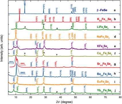
Figure 1. Powder X-ray diffraction patterns for samples measured at 297 K, Cu Kα radiation.
(a) β-FeSe; (b) K0.8Fe1.7Se2 (by high-temperature route); (c) Nominal LiFe2Se2; (d) Nominal NaFe2Se2; (e) Nominal KFe2Se2 (background corrected), peaks marked by ‘ ’ are due to unknown phase; (f) Nominal Ca0.5Fe2Se2; (g) Nominal Sr0.8Fe2Se2; (h) Nominal Ba0.8Fe2Se2; (i) Nominal EuFe2Se2; (j) Nominal Yb0.8Fe2Se2. Peaks marked by ‘*’ are due to residual β-FeSe.

