Fig. 5.
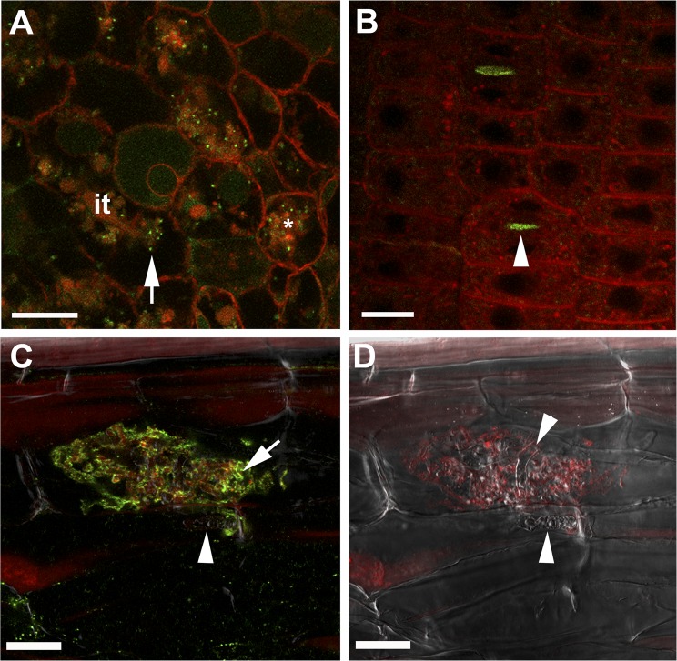
MtVAMP721e marked vesicles accumulate at the site of symbiosome, periarbuscular membrane formation and at cell plates. Confocal microscopy images showing localization of GFP-MtVAMP721e expressed under the control of its native promoter. (A) Accumulation of GFP-MtVAMP721e labeled dot-like structures (green) in infected Medicago nodule cells at local regions of infection threads (it) where bacteria are released (unwalled droplet, arrow) and around newly formed symbiosomes (*). (B) Accumulation of GFP-MtVAMP721e at the cell plate (arrowhead) in dividing meristematic cells of a Medicago root. (C and D) Confocal microscopy picture of immunolocalization of GFP-MtVAMP721e (C) and corresponding Bright-field image (D) in a root cortical cell containing an arbuscule. GFP was visualized by anti-GFP antibodies (green fluorescence). The signal is present near fine arbuscule branches (arrow) and absent on intraradical hypha (arrowhead). Samples were contrasted by FM4-64 (red) to visualize membranes. (Scale bars, 20 μm in A, C, and D; 10 μm in B).

