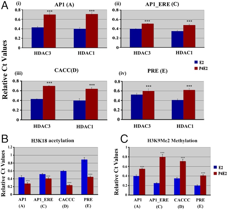
Fig. 4.
Acetylation and methylation signature on Mcm2 chromatin 3 h after hormone treatment. Uterine tissues were collected from ovariectomized mice after 3 h of E2 or P4E2 treatment and subjected to ChIP using antibodies against (A) HDAC1 or HDAC3, as described in the Materials and Methods. The results presented are compared to input and normalized against IgG control. Each site is independently shown. Differential binding as indicated ***P < 0.001, n = 5. (B) H3K18Ac levels at different sites were determined by Chip-QPCR 3 h after E2 or P4E2 treated uterine chromatin. (C) Levels of H3K9Me2 on different sites were determined by Chip-QPCR 3 h after E2 or P4E2 treated uterine chromatin. Statistical analysis (B and C) represents the difference between E2 treated samples with P4E2 treated one at four different sites, ***P < 0.001, n = 5.

