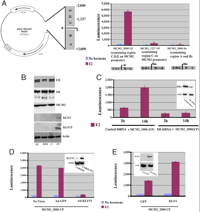
Fig. 5.
Transcriptional activity of the Mcm2 promoter in Ishikawa cells. (A) Shown is the representative diagram of PGL4 (luc2) vector containing the putative Mcm2 promoter region containing the specific sites for binding of transcription factors identified in Fig. 2. Ishikawa cells were transiently cotransfected with a control renilla plasmid and the luciferase derivatives containing three separate genomic fragments 2000 bp upstream region of TSS (MCM2_2000 UP), downstream region containing 2000 bp downstream in relation to TSS (MCM2_2000 DO), and region containing 1227 upstream in relation to TSS (MCM2_1227 UP) for 36 h followed by vehicle alone (blue bar) or E2 (red bar) treatment and assessed for luciferase activity 14 h after hormone treatment. Results in all panels are presented as normalized by firefly luciferase activity compared to Renilla control vector. (B) Serum deprived Ishikawa cells were treated with E2 or P4E2 and whole cell lysates were collected and analyzed by western blotting for expression of ERα, PR, MCM2, KLF4 and KLF15. The vehicle treated cells (C) along with uterine epithelial tissue lysates (CT) are used as control for KLF4 and 15, respectively. (C) Effects of silencing of ERα expression on Mcm2 transactivation. Ishikawa cells were cotransfected with ERα SiRNA along with the MCM2_2000(UP) construct followed by E2 treatment for 3 h or 14 h when the cells were harvested as described in the Materials and Methods. Insert is the Western blot analysis of ERα expression compared to actin control after transfection of specific SiRNA or scrambled SiRNA in Ishikawa at the end of the experiment. (D) Role of KLF15 in MCM2_2000 transactivation. Ishikawa cells were transfected with MCM2_2000 UP followed by infection with AdKLF15 or Ad-GFP. Cells were incubated for 36 h, then treated with E2 or vehicle for 14 h. Luciferase assays were performed on the isolated cells extracts. Insert is the Western blot analysis of KLF15 expression after infection with AdGFP (control) or AdKLF15 (experiment) compared to actin control in Ishikawa followed by E2 treatment for 14 h. (E) Role of KLF4 in MCM2_2000 transactivation. Ishikawa cells were cotransfected with MCM2_2000 UP along with KLF4 or vector containing GFP. Cells were incubated for 36 h then treated with E2 or vehicle for 14 h. Luciferase assays were performed on the cells extracts. Insert is the Western blot analysis of KLF4 expression compared to actin control after transfection of specific pGL3KLF4 (experiment) or pGL3GFP (control) in Ishikawa followed by E2 treatment for 14 h.

