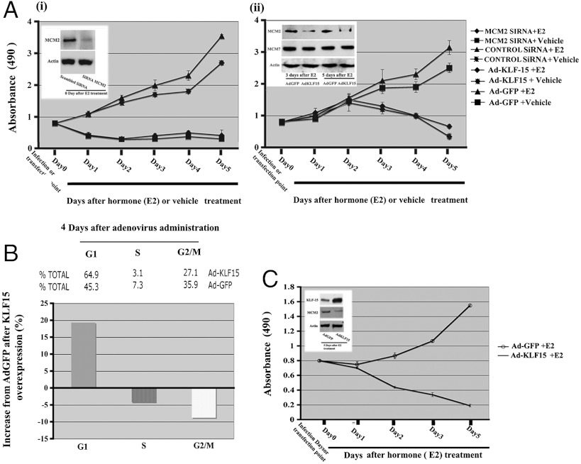
Fig. 6.
Effects of MCM2 knock down and KLF15 expression on Ishikawa cell proliferation. (A) Ishikawa cells were cultured in hormone free media for 24 h after MCM2 or scrambled SiRNA transfection (i) or infection with Ad-KLF15 or Ad-GFP (ii) followed by E2 or vehicle (ethanol) treatment. Cell proliferation was assayed in triplicate wells daily for 5 d after hormone treatment as described in the Materials and Methods, using the MTT assay (absorbance at 490 nm). Inserts (i, ii) indicate the Western blot analysis of equivalent amounts of protein isolated from these cell and detected with antibodies against MCM2, MCM7 (only ii) or actin as control after silencing of MCM2 SiRNA (i) or after KLF-15 overexpression (ii) for the indicated times. (B) Role of KLF15 overexpression on cell cycle parameters. Four days after postinfection, Ishikawa cells were collected and analyzed by flow cytometry to determine distribution of cells in each phase of the cell cycle. (C) Role of KLF15 overexpression on T47D cell proliferation. Human breast cancer T47D cells in culture were infected with rAdKLF-15 or rAd-GFP. Twenty-four hours after infection, cells were treated with E2 or vehicle and cell proliferation was assessed by the MTT method from 0 d to 5 d after treatment. Insert shows the Western blot analysis using antibodies against KLF15 and MCM2 compared to the control actin 5 d after treatment, indicating the reciprocal expression of KLF15 and MCM2. Values in (A) and (C) are presented as mean ± SE of five independent experiments (A and C).

