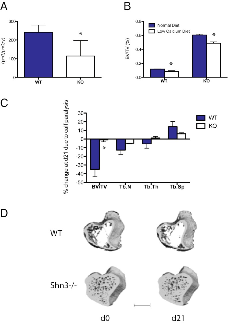
Fig. 4.
In vivo analysis of Shn3-deficient mice challenged with resorptive stimuli. (A) Dynamic histomorphometry was performed to quantify bone formation rate in 12-wk-old WT and Shn3−/− (KO) animals (n = 5 mice per group). *P < 0.05. (B) Eleven-week-old WT and Shn3−/− (KO) mice were fed a normal or low-calcium diet for 2 wk. BV/TV in the distal femoral metaphysis was determined by micro computed tomography (μCT) (n = 5 mice per group). (C) Six-month-old WT (n = 6) or Shn3−/− (KO, n = 8) mice were injected with botulinum toxin in the calf muscle. At day 0 (just before toxin injection) and day 21, the indicated parameters were determined by μCT at metaphyseal region of the proximal tibia. Data are expressed as percent change of the indicated parameter attributable to calf paralysis. Tb.N, trabecular number; Tb.Sp, trabecular spacing; Tb.Th, trabecular thickness. (D) Representative μCT images depicting WT and Shn3−/− samples at the level of the proximal tibia during the 21-d study. WT refers to the Shn3 fl/fl genotype, whereas cKO (conditional KO) refers to Shn3 fl/fl × Prx1-Cre animals. (Scale bar: 1 mm.)

