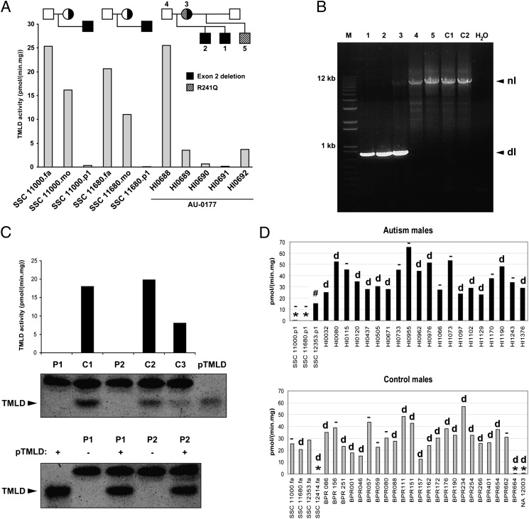
Fig. 3.
Genetic and enzymatic characterization of hemizygous deletion of exon 2. (A) TMLD activity measured in lymphoblast homogenates of three families with exon 2 deletion. (B) PCR assay results for the AU 0177 family showing the deletion in the two affected brothers (1, 2) and in the mother (3) but not in the father (4), unaffected maternal half-brother (5), or unaffected controls (C1 and C2). There is bias of amplification in the mother, such that the normal band is faint. dl, deletion; nl, normal. (C) (Upper) TMLD activity and Western blot analysis of 2 individuals with exon 2 deletion (P1, HI0690; P2, BPR664) and three controls (C1–C3). Purified TMLD (pTMLD) is used as a positive control. (Lower) Western blot analysis of 2 individuals (P1 and P2) with (+) or without (−) addition of pTMLD, showing the complete absence of protein in cases of exon 2 deletion and confirmation of the identity of the immunoreactive material as TMLD. The upper band in the Western blot is an irrelevant protein. (D) (Upper) TMLD activity measured in lymphoblast homogenates from several autism males. *,TMLHE exon 2 deletion; #, E287K; d, deletion in intron 1 in 13 individuals; −, no deletion in intron 1 in 9 individuals. SSC 12353.p1 was not tested for the presence of intron 1 deletion. (Lower) TMLD activity measured in lymphoblast homogenates from male controls. BPR indicates local unaffected controls, and NA 12003 is an unaffected individual. SSC 12353.fa was not tested for presence of intron 1 deletion. There was no apparent correlation of the level of enzyme activity with the presence or absence of the intronic deletion. For A, C, and D, assays were run in duplicate and the average is plotted without error bars. fa, father; mo, mother; p1, proband.

