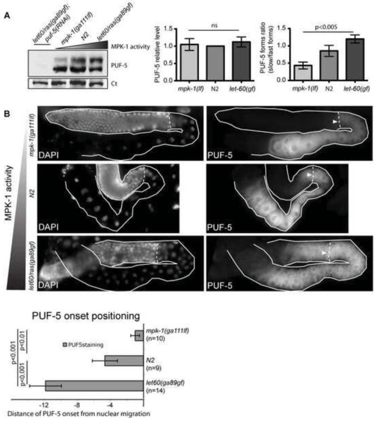
Fig. 8. MPK-1 controls PUF-5 protein forms and onset of expression.

(A) PUF-5 western blot: protein was extracted from equal numbers of synchronized adult hermaphrodites after 12 h at restrictive temperature for the indicated genotypes. Two PUF-5 forms were observed, which were strongly reduced after puf-5(RNAi). In three experiments, the ratio of slower/faster PUF-5 forms increased with Ras-MAPK activity. Bar graphs show quantification of total PUF-5 levels (left graph) normalized to loading control (Ct) band in the same blot, and ratio of slow/fast forms (right graph) by densitometry; ns=not signficant. (B) MPK-1 controls PUF-5 onset. Dissected gonads of indicated genotypes were stained in parallel for PUF-5, and imaged with matched exposures. DAPI staining revealed nuclear positioning. Bottom panel: Quantification of PUF-5 onset relative to nuclear migration from periphery to gonad core. Values are nuclear diameters from this transition (negative values are distal to transition). Error bars are s.d. for one representative experiment.
