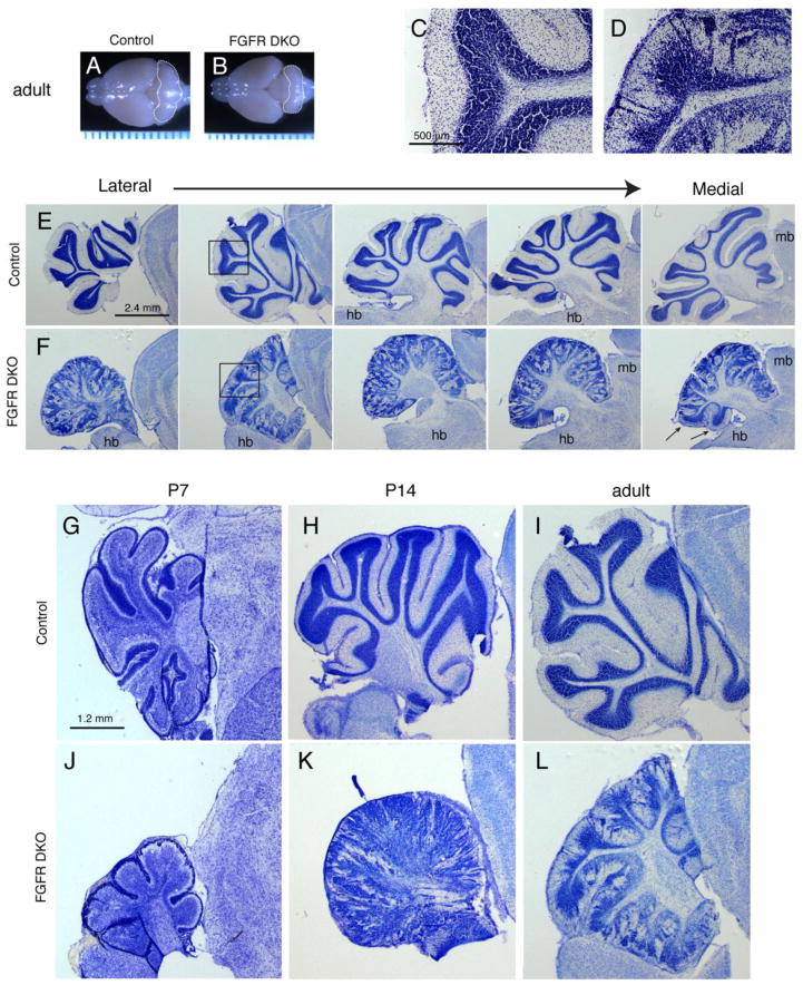
Figure 2. Fgfr1 and Fgfr2 Double mutants show decreased cerebellar volume and abnormal cerebellar lobule and layer morphology.
Whole mount view of adult (3 month old) brains of control (A) and FGF DKO (B) mice demonstrate the decreased size of the FGFR DKO cerebellum (outlined). Ruler units are in mm. Cresyl violet staining of sagittal sections from adult control (C,E) and FGFR DKO (D,F) mice demonstrate the abnormal laminar formation (compare C and D), and the malformed cerebellar cortex of FGFR DKO adults in the lateral to medial extent (E,F). The disruption occurs in a gradient with the most lateral sections and anterior lobules most disrupted. Arrows indicate posterior lobules on a medial section, which are spared from significant disruption. Developmental analysis of postnatal cerebellar morphology from P7 (G and J), P14 (H and K), and adult (I and L) control (G–I) and FGFR DKO (J–L) mice. Hindbrain, hb; midbrain, mb; ml, molecular layer; igl, internal granular layer.

