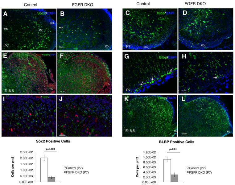
Figure 5. The severely disrupted cerebellum in adult FGFR DKO mice is a result of decreased number and altered radial orientation of glial cells.
Immunofluorescence staining for Sox2 at P7 in Control (A) and FGFR DKO mice (B). FGFR DKO had significantly fewer Sox2+ cells than control littermates (M). Immunofluorescence staining for BLBP in control (C,G) and FGFR DKO mice (D,H) at P7 demonstrates that in FGFR DKO animals (H) the Bergmann glia are significantly reduced in number (N), their cell bodies are further inside from the EGL than in the control, and there is a relative lack of radial glial fibers traversing the EGL and forming glial end feet attachments (double headed arrows in G and H) compared to controls. Sox2 and Reelin immunostaining of control (E,I) and FGFR DKO (F,J) embryos at E16.5 demonstrate a mild decrease in both Sox2 positive and Reelin positive cell bodies. Squares in E and F indicate location of high power images in I and J. BLBP staining in E16.5 embryos of control (K) and FGFR DKO (L) mice demonstrate that the defects in Bergmann glia number and morphology arise embryonically.

