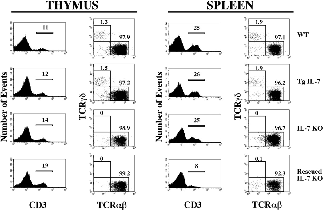
Figure 5. T cell lymphopoiesis remains deficient in IL-7 KO mice after selective expression of IL-7 by osteoblasts.
Cell suspensions from thymus and spleen from WT, Tg IL-7, IL-7 KO and rescued IL-7 KO mice were stained with anti-CD3, anti-TCRαβ and anti-TCR-γδ antibodies coupled to distinct fluorochromes, and analyzed by flow cytometry as described. Histograms on the left showed the percentage of cells reactive to anti-CD3 and plots in the right show the distribution of TCRαβ and TCRγδ within CD3 gated populations.

