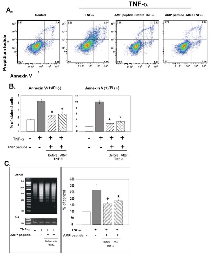
Fig. 4.

Antiapoptotic effects of antrum mucosal protein peptide (AMP-p) in human adult low-calcium, high-temperature keratinocyte (HaCaT) cells exposed to tumor necrosis factor (TNF) α. HaCaT cells in Dulbecco's modified Eagle's medium (DMEM) plus 0.5% fetal bovine serum (FBS) were exposed to TNF-α (50 ng/mL) in the presence or absence of AMP-p (8 μg/mL, 2 h before or after TNF-α). Control cells were untreated. Apoptosis was assessed by flow cytometry of annexin V– and propidium iodide (PI)–stained cells. A, Data from a single representative experiment. B, AMP-p inhibited the TNF-α–induced increase in annexin V-positive/PI-negative cells (left) and annexin V-positive/PI-positive cells (right). Values are means ± standard errors for four independent experiments. Asterisk, p < 0.001. C, DNA laddering was assessed by ligase-mediated (LM) polymerase chain reaction (PCR) to evaluate apoptosis as described in Fig. 3C. Left, LM-PCR products were resolved on agarose gel. Right, LM-PCR results were quantified and normalized to the En-2 bands. Values are percent of control apoptosis (100%) shown as mean ± standard error for four experiments. Asterisk, p < 0.001.
