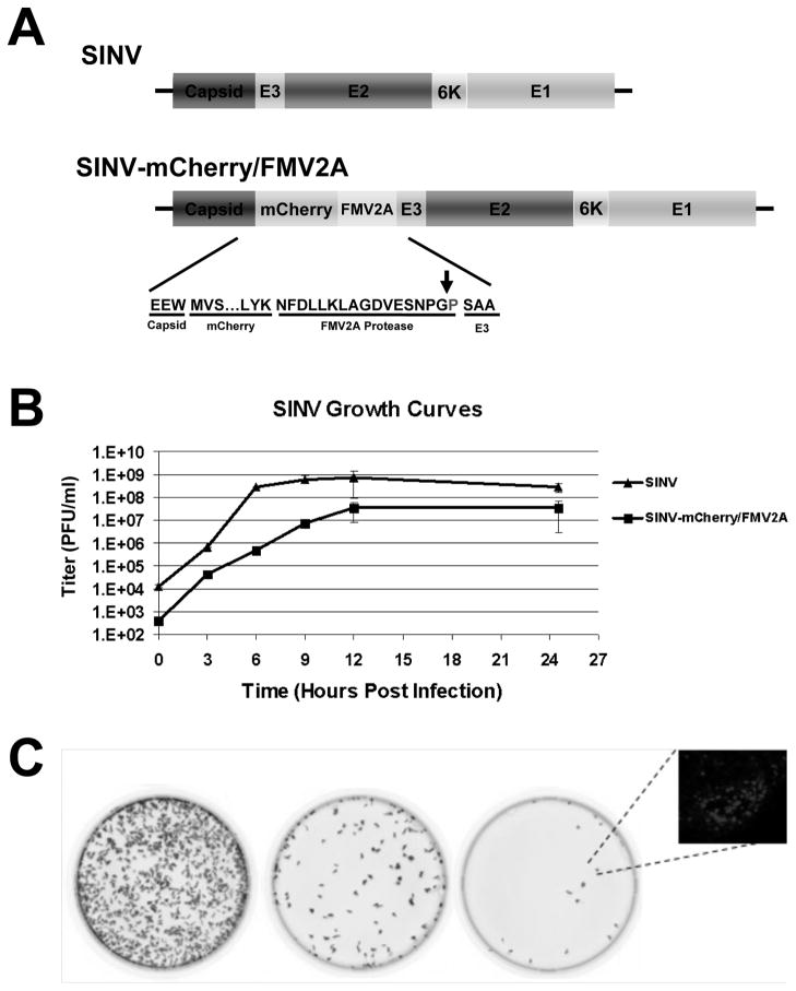
Figure 1. SINV-mCherry/FMV2A Produces Fluorescent Foci Upon Infection of Tissue Culture Cells.
A) Schematic of the structural ORF of wild-type SINV and SINV-mCherry/FMV2A. Inset is the primary amino acid sequence of the cleavage site of the FMV2A protease and the arrow indicates the site of cleavage. B) Single-step growth curve of SINV-mCherry/FMV2A and wild-type SINV derived from BHK-21 cells and titered on BHK cells. Error bars indicate the standard deviation of three independent replicates. C) Phosphorimager scans of confluent BHK-21 monolayers infected with 10-fold dilutions of SINV-mCherry/FMV2A. Inset is a fluorescent microscopy image of a representative BHK-21 fluorescent focus at 24 hours post infection under 40x magnification.

