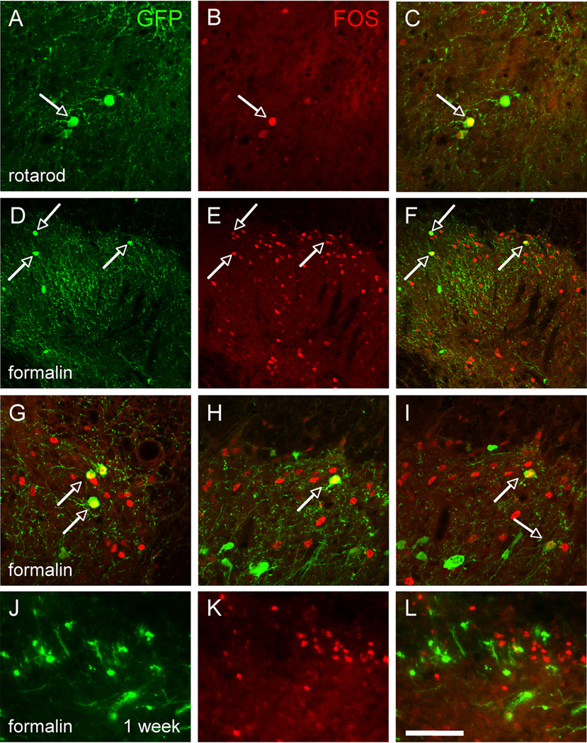
Figure 5. Transplanted MGE-derived neurons are activated by innocuous and noxious peripheral stimulation.
(A–C) Walking for 90’ on a rotarod induced expression of Fos (red) in MGE cells, indicating that innocuous peripheral stimuli (in this case conveyed by myelinated primary afferents) activate the MGE transplants. (D–L) Hindpaw injection of formalin (1%) also induces expression of Fos (red) in a large number of spinal cord neurons, including GFP+ (green) MGE-derived neurons, 1 month after transplantation (D–I) but not at 1 week post-transplantation (J–L). Arrows point to examples of Fos and GFP+ double-labeled cells. Scale bars equals 50 µm in A–C and G–L, 100 µm in D–F.

