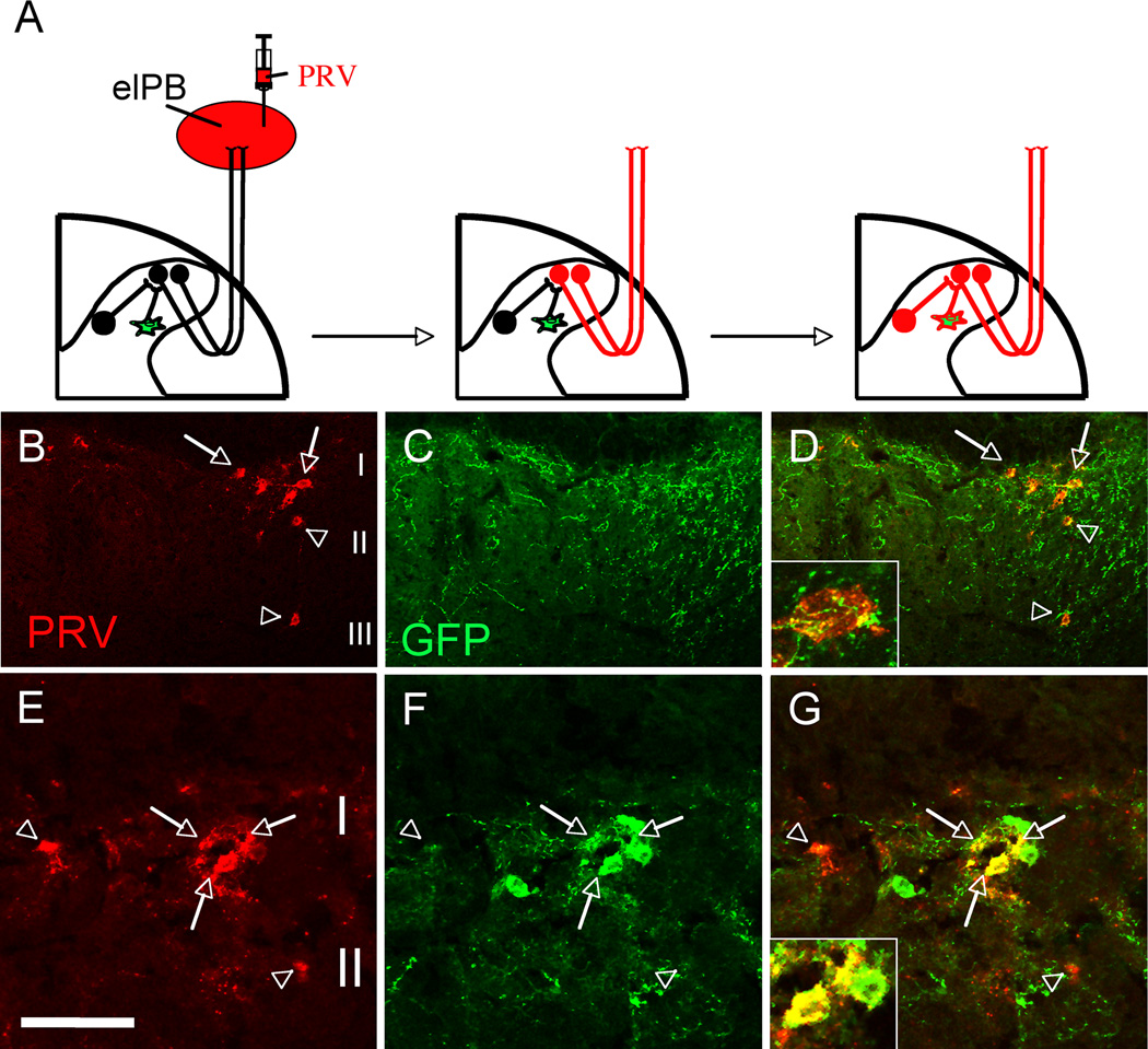
Figure 7. Transplanted MGE-derived neurons integrate into spinal cord circuits that engage lamina I projection neurons.
(A) Injection of the retrograde transneuronal viral tracer pseudorabies (PRV, red) in the external lateral parabrachial nucleus (elPB; left panel) results in infection of spinal cord projection neurons that target the elPB (projection neurons “become” red; middle panel). Subsequent retrograde transfer of PRV labels spinal cord neurons that are upstream of the infected projection neurons (spinal neurons, including GFP+ (green) MGE cell “become” red; right panel). (B–D) PRV+ (red) projection neurons of spinal cord lamina I (arrows) and PRV+ interneurons of laminae II–III (arrowheads). Note the extensive arborization of GFP-positive terminals around retrogradely PRV-labeled neurons (D, inset; see also Supplementary Figure 5). (E–G) MGE transplanted cells (green) are among the cells that are retrogradely infected by PRV (red), indicating that MGE cells have integrated into a neuronal circuit that influences projection neurons of lamina I. Arrowheads in E point to retrogradely infected PRV+/GFP− spinal cord interneurons. Arrows point to examples of PRV+/GFP+ double-labeled cells. Arrowheads Scale bars equals 100 µm in B–D and 50 µm in E–G.

