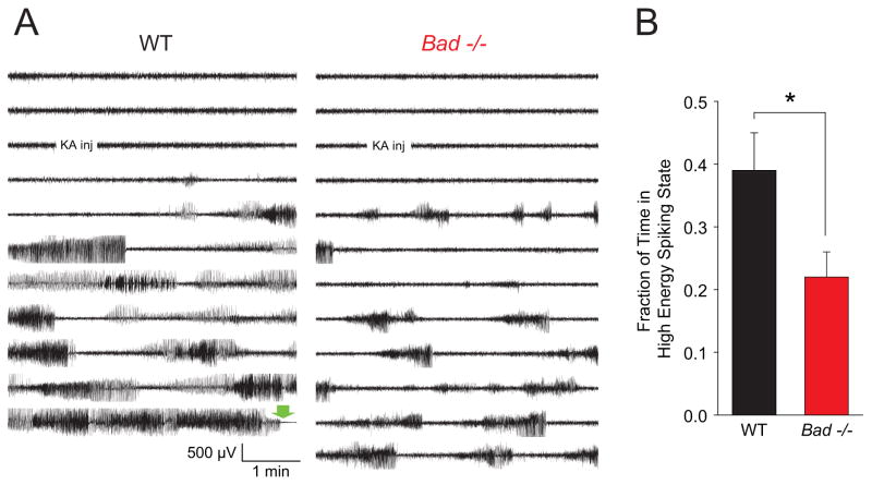
Figure 4. EEGs of Kainic Acid-Induced Seizure Activity in Wild-Type and Bad−/− Animals.
(A) A single representative EEG channel recording of approximately 1 hour for each genotype (right frontoparietal electrode). A single i.p. injection of kainic acid (30 mg/kg of body weight) was administered at the indicated time. The green block arrow near the end of the wild-type recording indicates the time of death during generalized tonic-clonic status epilepticus. (B) The average fraction of time spent in a high energy spiking period for each genotype during a 30 minute period beginning 3 minutes after injection. EEGs were scored by an investigator blind to the genotype, guided by both the raw EEG traces and a trace of power in the 20–70 Hz band to identify onset and offset of high energy spiking (typically seen as rapid increases or decreases of ≥5 dB in power). Wild-type, n=17; Bad−/−, n=20; mean ± SEM, * p < 0.05 by two-tailed Student’s t-test. In these cohorts, 9 of 17 wild-type and 0 of 20 Bad−/− animals died during status epilepticus.

