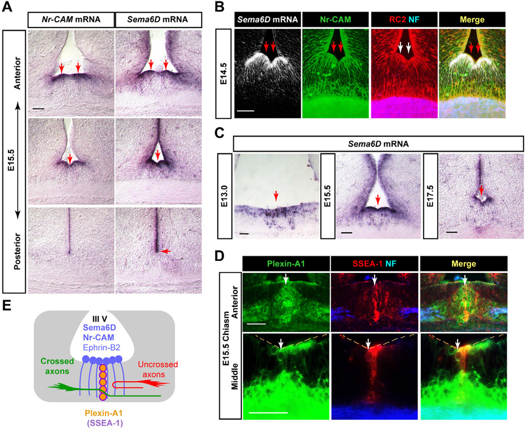
Figure 1. Expression of Sema6D and Plexin-A1 at the optic chiasm.
(A) Sema6D and Nr-CAM are expressed in radial glia at the chiasm midline at E15.5, but Sema6D extends further dorsally in the VZ. (B) Sema6D mRNA is expressed in Nr-CAM+ (green)/RC2+ (red) radial glia at E14.5. Retinal axons stained with neurofilament antibody (NF, blue) extend through radial glial processes. (C) Sema6D mRNA is expressed in radial glial cell bodies at the floor of the third ventricle in the optic chiasm and along the ventricular zone at E13.0, E15.5 and E17.5. (D) Plexin-A1 expression at the E15.5 optic chiasm in cells in the ventral diencephalon. In frontal sections, Plexin-A1 protein (green) is colocalized with SSEA-1 (red) in chiasmatic neurons at the midline raphe and also on neurofilament+ retinal axons (blue). (E) Schema of Sema6D and Plexin-A1 expression at the optic chiasm, depicted in frontal planes. Note that Sema6D is expressed by Nr-CAM+/ephrin-B2+ radial glial cells, and Plexin-A1 is expressed by SSEA-1+ chiasm neurons. Scale bars: 100µm.

