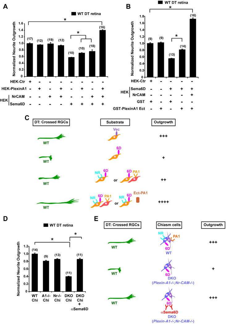
Figure 3. Nr-CAM and Plexin-A1 in cells of the optic chiasm convert the inhibitory effect of Sema6D on crossed RGCs to growth-promotion.
(A) Quantification of WT DT retinal outgrowth on Sema6D+/Nr-CAM+ HEK cells and/or Plexin-A1+ HEK cells. Note that inhibition of DT outgrowth by Sema6D is partially alleviated by Plexin-A1+ or by Nr-CAM+ HEK cells. However, DT neurite outgrowth is greatly enhanced (by ~40%) in the presence of Sema6D+/Nr-CAM+ HEK cells cocultured with Plexin-A1+ HEK cells. (B) Quantification of WT DT retinal outgrowth on Sema6D+/Nr-CAM+ HEK cells in the presence of 100µg/ml GST-Plexin-A1 ectodomain or GST proteins. DT RGC outgrowth is poor on Sema6D+ HEK cells, but inhibition is partially alleviated by the addition of GST-Plexin-A1. However, DT neurite outgrowth on Sema6D+/Nr-CAM+ HEK cells is greatly enhanced (by ~70%) in the presence of GST-Plexin-A1. (n) = number of explants for each condition, * p<0.01 (C) Summary of results in Figures 3A and B. Note that outgrowth of crossed RGC axons is enhanced on Sema6D+/Nr-CAM+ HEK cells cocultured with Plexin-A1+ HEK cells and especially by GST-Plexin-A1 ectodomain. (D) Quantification of WT DT retinal outgrowth on Plexin-A1−/− (A1−/−), Nr-CAM−/− (Nr−/−) single mutant or Plexin-A1−/−;Nr-CAM−/− (DKO) double mutant chiasm cells. (E) Summary of results in Figure 3D. Note that DKO chiasm cells are unsupportive of WT RGC neurite outgrowth, but with αSema6D DT neurite outgrowth is partially restored. (n) = number of explants for each condition, * p<0.01

