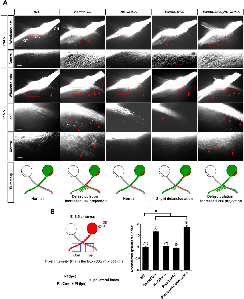
Figure 7. Sema6D, Nr-CAM and Plexin-A1 are important for formation of the crossed RGC projection in vivo.
(A) Whole mounts of the optic chiasm in E14.5 and E15.5 WT, Sema6D−/−, Nr-CAM−/−, Plexin-A1−/− or Plexin-A1−/−;Nr-CAM−/− mutants unilaterally labeled with DiI in the retina at the optic disc. E14.5 and E15.5 both Nr-CAM−/− and Plexin-A1−/− mutant chiasm have a normal ipsilateral projection (arrows) and only slight defasciculation of the contralateral projection (arrowhead) at E14.5. In contrast, in E14.5 Sema6D−/− and Plexin-A1−/−;Nr-CAM−/− mutant chiasms, RGC axons on the side of the chiasm contralateral to the midline are defasciculated (arrowheads); at E15.5 both ipsi- and contralateral projections in these mutants are highly defasciculated (arrowheads) and the ipsilateral projection is greatly increased (arrows). (n ≥ 3 embryos for each condition). Schematic depicts a summary of WT and mutant chiasm phenotypes. (B) Quantification of decussation defects in E15.5 Sema6D−/− single and Plexin-A1−/−;Nr-CAM−/− double mutant embryos. Schematic presentation shows the measurement of pixel intensities of contralateral and ipsilateral optic tracts and the calculation to obtain an ipsilateral index. Sema6D−/− and Plexin-A1−/−;Nr-CAM−/− mutants display a 1.6–1.9 times larger ipsilateral projection compared to WT, Nr-CAM−/− and Plexin-A1−/− chiasm. ON, optic nerve; OT, optic tract; OC, optic chiasm. Scale bars, 200µm in low-magnification images; 20µm in high-magnification images, (n) = number of explants for each condition, * p<0.01.

