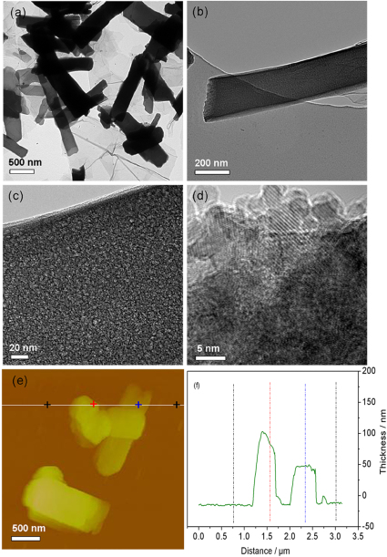
Figure 2. Morphological and structural analysis of iron glycolate ribbons after growth for 5 h.
(a–c) Typical TEM images with different magnifications. (d) HRTEM image reveal the monodispersed ribbons with porous architectures. (f) AFM image and the corresponding thickness analysis taken around the white line in (f) reveals a uniform thickness of about 50 nm.

