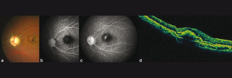
Figure 1.

Active CNV as seen on clinical photography (CP) (a), FFA (b, c) and OCT (d). FFA shows an active subfoveal classic choroidal neovascular membrane with profuse leakage in the late phases. Blocked choroidal fluorescence due to the overlying hemorrhage is also noted. OCT shows subfoveal CNVM and sub-retinal fluid
