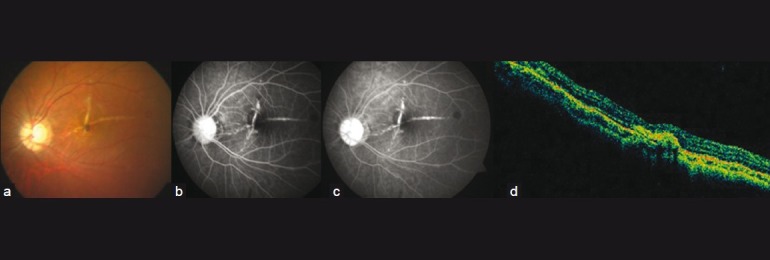
Figure 3.

Color fundus photograph (a), FFA (b, c) and OCT (d) at 6 months’ (final) follow-up. Fundus shows two lacquer cracks almost perpendicular to each other. FFA shows absence of leakage. OCT shows CNV regression with scarring and resolution of sub-retinal fluid
