Figure 1.

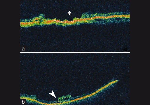
(a and b) Optical coherence tomography scan through pigmented lattice degeneration demonstrating irregularly thinned retina and full thickness defects (asterisk). (b) Full thickness hole at the edge of the lesion (arrowhead)

(a and b) Optical coherence tomography scan through pigmented lattice degeneration demonstrating irregularly thinned retina and full thickness defects (asterisk). (b) Full thickness hole at the edge of the lesion (arrowhead)