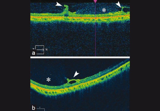
Figure 2.

(a and b) Optical coherence tomography image of non-pigmented lattice degeneration demonstrating triangular attachment of vitreous (arrowheads) and dense hyper-reflective dots over lesion (asterisk). The retinal thickness within the lesion is similar to adjoining retina
