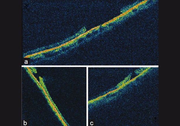
Figure 3.

(a–c) Optical coherence tomography images of peripheral retinal holes demonstrating similarity to idiopathic macular holes. (a) Flattened edges of the hole. (b) and (c) Everted edges, vitreous attachment and cystoid changes in surrounding retina
