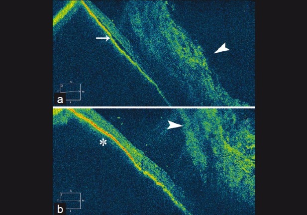
Figure 7.

Postoperative optical coherence tomography (OCT) images of patient who underwent scleral buckling for inferior retinal detachment and vitreous hemorrhage following trauma. There was minimal subretinal fluid (SRF) seen hazily through the hemorrhage running toward the break in the immediate postoperative period. (a) OCT scan through the area showing SRF (line arrow) and overlying vitreous hemorrhage (arrowhead); (b) Beyond the region shown in (a), no subretinal fluid was seen. A gentle indentation of the buckle can be made out (asterisk) with overlying vitreous hemorrhage (arrowhead)
