
Fig. 2.
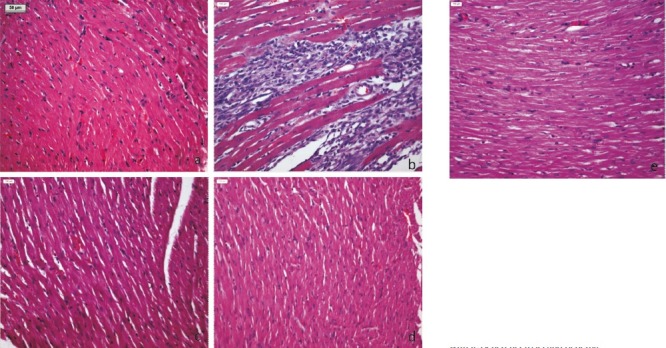
Light micrograph (H&Ex100) of rat's myocardium (a) Sham group showing normal histoarchitecture of myocardium, (b) I-R control group showing confluent necrosis of myofibrils, edema and infiltration of inflammatory cells with extravasations of red blood cells, (c) Benazepril 30 mg/kg showing reduced myonecrosis, oedema and infiltration of inflammatory cells, (d) A. paniculata 200 mg/kg pretreatment showing normal histoarchitecture of myocardium and (e) A. paniculata 200 mg/kg + I-R group showing resemblance to normal myocardial histoarchitecture with lessened necrosis and oedema.