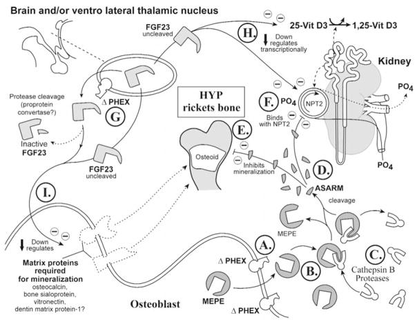
Figure 4.
X-linked rickets (Hyp). A cartoon illustrating the proposed changes in X-linked hypophosphatemic rickets. The primary defect in this disease is the PHEX gene, and this has an impact on renal phosphate handling, vitamin D metabolism, and mineralization. For comparison with normal states, see Fig. 3. See text for detailed description (specifically, the subparagraph entitled ‘X-linked hypophosphatemic rickets [Hyp] [Global Hypothesis]’). Also refer to the Table for more details concerning the icons used to represent the diverse molecules, pathways, and tissues.

