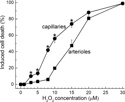
Fig. 1.
Induced cell death in capillaries and the precapillary tertiary arterioles in retinal microvascular complexes exposed for 20 h to various concentrations of H2O2. For each point, 10.9 ± 0.5 microvessel-containing coverslips were assayed. *H2O2-induced cell death was significantly greater in capillaries than in tertiary arterioles; the P values comparing capillary and arteriolar cell death induced by 3, 5, 8, 10, and 15 μM H2O2 were 0.014, 0.007, <0.001, <0.001, and 0.003, respectively.

