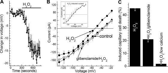
Fig. 2.
KATP channel activation in retinal microvessels during exposure to H2O2. A: time course for the hyperpolarizing effect of 15 μM H2O2. Values are the means of 4 experiments. During H2O2 exposure, the membrane potential increased significantly (P < 0.001). B: during continuous recordings (n = 4), current-voltage (I-V) plots were generated during exposure of the retinal microvascular complex to solution A (control, ■), 9 ± 2 min after the onset of exposure to solution A supplemented with 15 μM H2O2 (●), and finally 11 ± 2 min after the addition of 0.5 μM glibenclamide to the H2O2-containing solution (○). Exposure to H2O2 significantly (P = 0.001, paired t-test) increased the ionic conductance; glibenclamide significantly (P = 0.001, paired t-test) decreased the H2O2-induced conductance. Inset: I-V relations of the H2O2-induced conductance, which is the difference between the control and H2O2 I-V plots shown in this panel. C: effect of glibenclamide and low extracellular calcium on H2O2-induced capillary cell death. Microvessels were exposed for 6 h to 15 μM H2O2 in solution A without additives (H2O2 group), in solution A with 0.5 μM glibenclamide, or in solution A lacking added calcium. For the H2O2, H2O2/glibenclamide, and H2O2/low calcium groups, 14, 5, and 6 microvessel-containing coverslips were assayed, respectively. H2O2-induced capillary cell death was significantly (*P < 0.001) less in the glibenclamide and low calcium groups.

