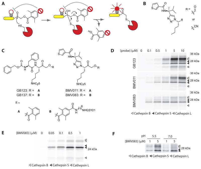
Figure 1.
Non-peptidic cysteine cathepsin activity-based probes. A) Schematic presentation of the mechanism of action of a quenched ABP. B) Structure of the cathepsin S selective aldehyde and nitrile inhibitors reported by the Ellman lab. C) Structures of the peptidic activity-based probes GB123 and the quenched GB137 and the non-peptidic probes BMV011 and the quenched BMV083. D) Labeling profile of GB123, BMV011 and BMV083 in living RAW cells. Cells were exposed to the indicated concentrations of probe for 3 hr, before being harvested, washed and lysed. 40 μg total protein was resolved on 15% SDS-PAGE and fluorescently labeled proteins were visualized by in-gel fluorescence scanning. E) Labeling profile of BMV083 in living human primary macrophages. Cells were exposed to the indicated concentrations of BMV083 for 3 hr, before being harvested, washed and lysed. 40 μg total protein was analyzed as described above. F) BMV083 labeling of RAW cell lysate (35 μg total protein) at pH 5.5 and 7.0 with indicated concentrations of probe for 1 hr. Labeled proteins were analyzed as described above. See also supplemental figures S1–S4.

