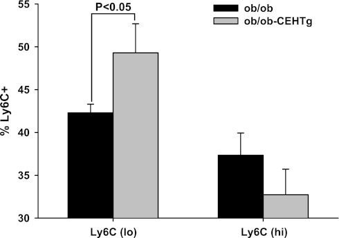
Fig. 8.
Increased number of Kupffer cells in Ly6Clo or M2 phenotype in ob/ob-CEHTg mice. Isolated kupffer cells were stained for leukocytes (CD45), macrophages (CD11b), and Ly6C and analyzed by FACS. The distribution of Ly6Clo and Ly6CHi is plotted as the %total CD45+CD11b+Ly6C+ cells. Data are presented as means ± SD; n = 5.

