Fig. 10.
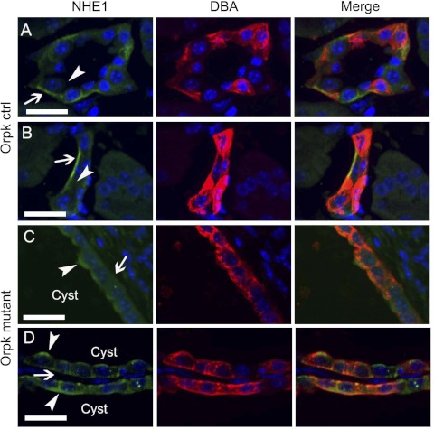
Localization of NHE1 in wild-type (WT) and Oak Ridge polycystic kidney (orpk) cystic kidneys. A and B: in 2 sets of representative confocal images from postnatal day 21 (P21) WT kidney sections, NHE1 (green) localized to the basolateral membrane (arrow) but not the apical membrane (arrowhead) of collecting ducts identified by DBA labeling (red). ctrl, Control. C and D: in two sets of representative confocal images from P21 Orpk cystic kidneys, NHE1 (green) localized to both the basolateral membrane (arrow) and the apical membrane (arrowhead) of collecting ducts identified by DBA labeling (red). In all panels, nuclei were counterstained with Hoechst (blue). “Cyst” refers to the lumen of a cyst. Scale bars, 25 μm.

