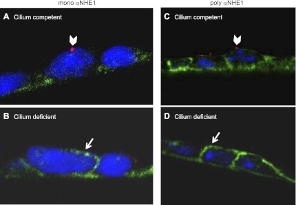
Fig. 8.
Localization of NHE1 in cilium-competent and -deficient cells. Monolayers grown on permeable supports were colabeled with either a monoclonal or a polyclonal antibody to NHE1 (mono αNHE1 or poly αNHE1, respectively) (in green), and antibodies to tubulin (in red). Folded membranes allowed for side-view imaging of the apical and basolateral membranes. A: a cilium-competent cell with a detyrosinated α-tubulin-labeled cilium (arrowhead) displayed predominantly basolateral expression of NHE1. B: a cilium-deficient cell displayed both apical (arrow) and basolateral expression of NHE1. Labeling was repeated in another experimental set. C: a cilium-competent cell with an acetylated tubulin-labeled cilium (arrowhead) displayed predominantly basolateral expression of NHE1. D: a cilium-deficient cell displayed both apical (arrow) and basolateral expression of NHE1. In all panels, DNA was stained with DAPI (in blue).

