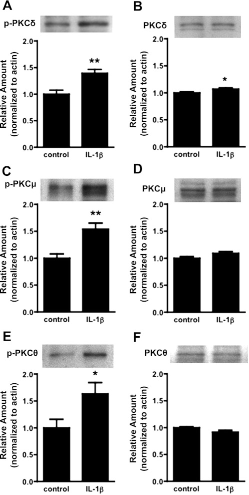
Fig. 3.
Activation of novel PKC isoforms in hBMECs in response to IL-1β exposure. Cells were treated for 1.5 h with IL-1β (100 ng/ml), and Western blots were performed with hBMEC lysates to probe for activation (phosphorylation) and expression of novel PKC isoforms δ, μ, and θ. There were significant increases in amounts of phosphorylated (p-)PKC δ, μ, and θ (A, C, and E). There were no substantial changes in expression of total PKC δ, μ, or θ (B, D, and F), although a modest increase in expression of PKC-δ was noted. Blots represent n ≥ 4 independent experiments, with quantitative data at bottom (band intensity normalized to β-actin). (*P < 0.05, **P < 0.01).

