Fig. 1.
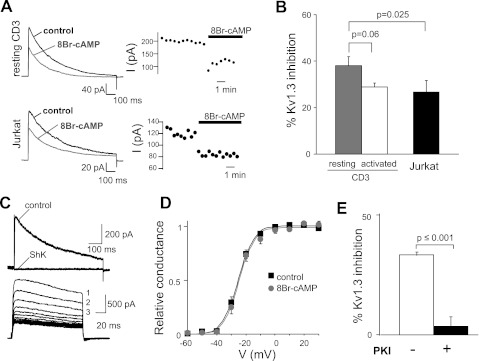
Activation of PKA significantly decreases KV1.3 activity. A: representative macroscopic KV1.3 currents (I) in resting CD3+ T cells (left top) and Jurkat cells (left bottom) before and after extracellular application of 8-bromoadenosine 3′,5′-cyclic monophosphate (8-BrcAMP; 1 mM). Corresponding changes in KV1.3 peak currents with time after addition of 8-BrcAMP are shown at right. Currents were elicited by depolarizing pulses to +50 mV from a holding potential (HP) of −80 mV every 30 s. B: average inhibition of KV1.3 currents by 8-BrcAMP in resting (n = 7) and activated (n = 10) CD3+ cells and in Jurkat (n = 10) cells. C: characteristic electrophysiological and pharmacological properties of KV1.3 currents in Jurkat T cells. Top: representative KV1.3 currents recorded before and after steady-state inhibition by ShK (10 nM). KV1.3 currents were elicited by depolarizing voltage steps to +50 mV from −80-mV HP every 30 s. Bottom: cumulative inactivation of KV1.3 channels. Cumulative inactivation was induced by consecutive depolarizing pulses to +50 mV from −80-mV HP applied every s. Current amplitude progressively decreased with each successive pulse (indicated by progressive numbers). D: average conductance (relative to the maximum conductance at +50 mV)-voltage curves for KV1.3 in resting CD3+ cells before (control) and after addition of 8-BrcAMP. Currents were elicited by depolarizing steps from −60 mV in 10-mV increment (−80-mV HP) every 30 s. E: average inhibition of KV1.3 currents by 8-BrcAMP in resting CD3+ cells with and without addition of 25 μM of the protein kinase inhibitor PKI6–22 in the pipette solution (n = 10 without PKI6–22 and n = 8 with PKI6–22).

