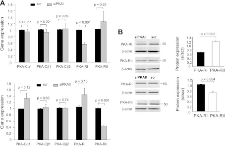
Fig. 2.
Specificity of small interfering (si)RNAs against PKA-RIα (siPKAI) and PKA-RIIα (siPKAII). A: siPKAI and siPKAII selectively downregulate the corresponding regulatory subunits but not the catalytic subunits. Gene expression of PKA catalytic (PKACα1, PKACβ1, and PKACβ2) and regulatory (PKA-RI and PKA-RII) subunits was determined by RT-quantitative (q)PCR in Jurkat cells 48 h after nucleofection with siRNA against PKA-RIα (siPKAI; top) and PKA-RIIα (siPKAII; bottom) or scrambled sequence siRNA (scr). Gene expression for the PKA subunits is represented as the fold change in PKA subunit gene compared with the housekeeping gene GAPDH. Data are the average of separate experiments each in quadruplicate: PKACα1 (n = 5), PKACβ1 (n = 4), PKACβ2 (n = 4), PKA-RI (n = 5), and PKA-RII (n = 3) in cells transfected with siPKAI. For siPKAII transfected cells, the expression of each PKA subunit was determined in 3 separate experiments. Data are normalized to scr controls (n = 6 for siPKAI and n = 3 for siPKAII). B: PKA-RI, but not PKAII, protein expression was downregulated by siPKAI. Left: immunoblotting of PKAI and PKAII in scr- and siPKAI-treated cells. Corresponding β-actin is shown at bottom. Predicted molecular weights for PKA-RI and PKA-RII are 48 and 51 kDa, respectively. Right: RI and RII protein levels (normalized to β-actin) are reported as relative to scr (n = 3; means ± SD). B: siPKAII selectively downregulates PKA-RII protein expression in Jurkat cells. Representative immunoblot of PKAI (top) and PKAII (bottom) in scr- and siPKAII-treated cells are shown at left. Corresponding β-actin is shown underneath. Average band intensities normalized to β-actin and relative to scr are reported at right (n = 3).

