
Figure 1.
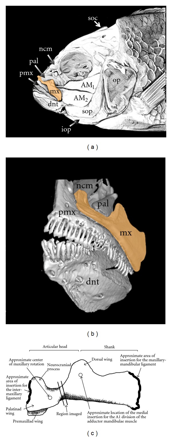
(a) Illustration of cichlid craniofacial anatomy in the lateral view. (b) Micro-CT scan of the oral jaws and associated elements. (c) Anatomy of a cichlid maxilla (left side, lateral view) showing the region imaged using μCT scanning. In panels (a) and (b) the maxilla (mx) is highlighted orange. Drawing by Kristen Ann Tietjen. AM1: first division of the adductor mandibulae muscle; AM2: second division of the adductor mandibulae; dnt: dentary; iop: interopercle; ncm: neurocranium; op: opercle; pal: pterygoid process of the palatine; pmx: premaxilla; soc: supraoccipital crest of the neurocranium; sop: subopercle.