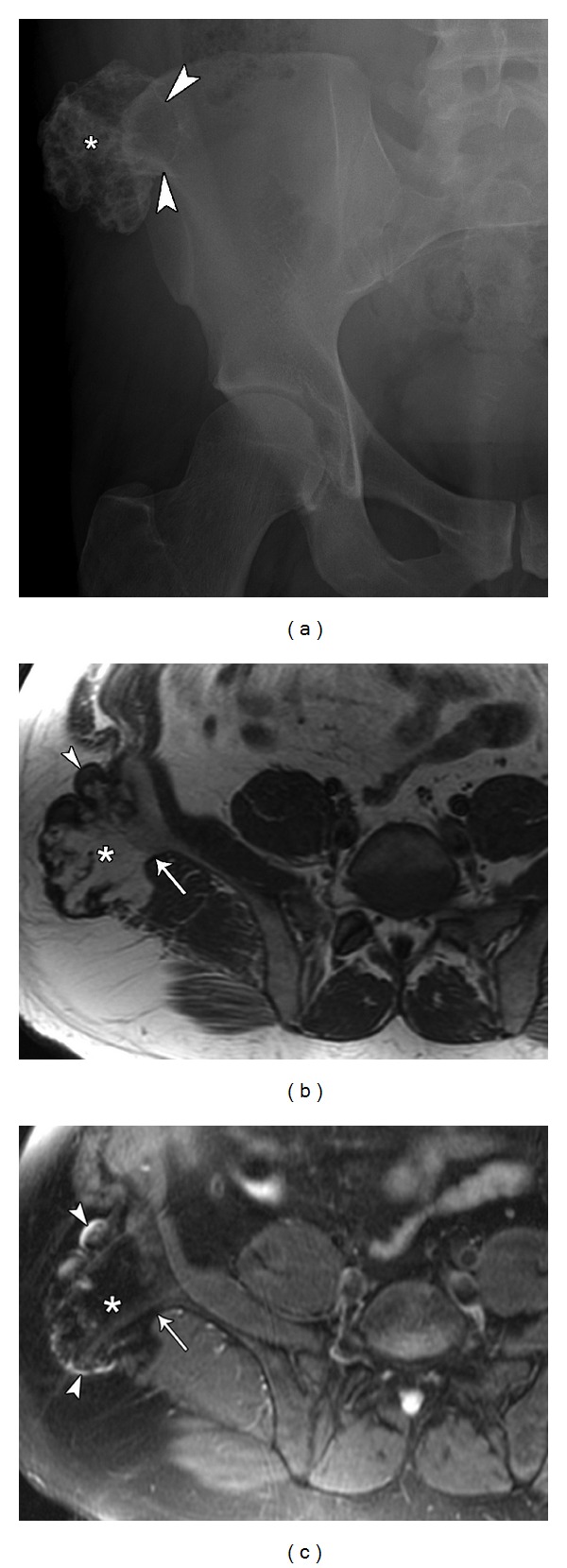
Figure 2.

31-year-old male presents with palpable hard mass lateral to right iliac crest diagnosed as an osteochondroma. Pelvic plain film (a) demonstrates an irregularly calcified, pedunculated lesion (asterisk) arising from the right iliac crest. It presents as a peripheral outgrowth with its cortex in continuity with the iliac bone (arrowheads). Axial T1 (b) and axial T2 fat sat. (c) MRI images of same lesion (asterisk) demonstrate the continuity of the cortex and medullary portion of the lesion with the parent bone (arrow) and identify a thin cartilage cap (arrowheads).
