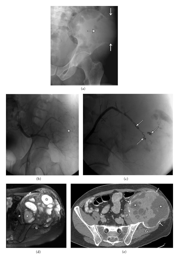
Figure 4.

40-year-old male with a large left iliac wing expansile mass diagnosed as pelvic giant cell tumor. Plain film (a) shows a large, lytic, destructive lesion in the right iliac wing with indistinct margins and cortical breakthrough (arrows), with no visible matrix. Left internal iliac artery angiogram (b) demonstrating the highly vascular nature of the tumor. Selective embolization of the superior gluteal artery (c) demonstrating embolization coils (arrowheads), and decreased tumor vascularity. Axial T2 fat sat MRI (d) and axial post-contrast CT images (e) demonstrating expansile nature of the giant cell tumor with multiple foci of necrosis (asterisk) and intervening tumor stroma. Size of the mass increased following embolization due to internal bleeding and necrosis. Displacement of surrounding soft tissues (Iliopsoas (P), gluteal musculature (G)), rather than invasion is illustrated. Note residual outer rim of expansile cortical bone (arrows), better appreciated on the CT scan.
