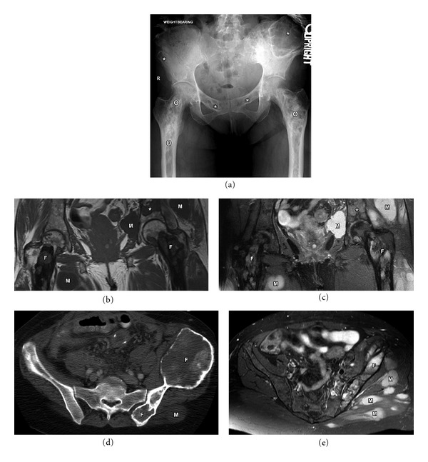
Figure 5.

61-year-old female with Mazabraud syndrome (polyostotic fibrous dysplasia and soft tissue myxomas (M)). Plain film (a) demonstrates multiple lucent lesions with ground glass matrix in the pelvis (asterisk) and long predominantly sclerotic abnormality in bilateral femurs (circle). Fibrous dysplasia is often described as a long lesion in the long bone. Coronal T1 (b) and STIR (c) MRI images demonstrating multiple fibrous dysplasia (F) lesions. Note that some of these maintain low to intermediate signal intensity on both T1 and T2 images (asterisk). Axial CT (d) and axial T2 fat sat (e) MRI images demonstrating contour abnormality of the left ilium with ground glass matrix and focal bone expansion (F). Multiple T2 bright myxomas (M) are noted in the gluteal musculature.
