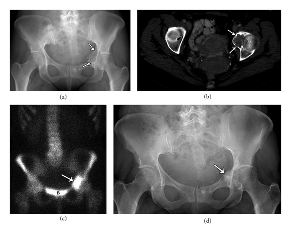
Figure 7.

Forty-six-year-old female with a lytic lesion in the left medial acetabulum, diagnosed as chondroblastoma. Plain film (a) and axial CT (b) demonstrating a lytic lesion with heterogeneous chondroid matrix and sharp scalloped margins. The tumor showed increased uptake on bone scan (c) (B: bladder). Plain film (d) demonstrating healed chondroblastoma following surgical curettage and cement packing.
