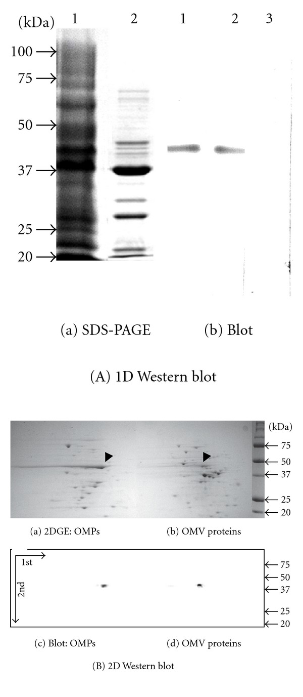
Figure 3.

EF-Tu detected in OMV and OM fractions. Panel (A): (a) SDS-PAGE of proteins from OM (lane 1) and OMV fractions (2). (b) Western blot of proteins from OM (lane 1) and OMV fractions (2) reacted with the EF-Tu antibody diluted at 1 : 3000. Control blot with pre-immune serum (lane 3). Panel (B): 2D-based Western blot. Proteins from OM (a and c) and OMV fractions (b and d) were resolved by isoelectric focusing (1st D) and then separated on a second dimension SDS-PAGE (2nd D). Proteins from the gel were blotted onto PVDF membranes and probed with the anti-EF-Tu antibodies (c and d). Arrows: EF-Tu. Blot with pre-immune serum not shown.
