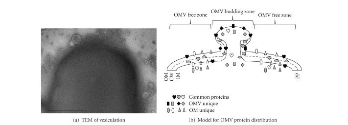
Figure 6.
EF-Tu delivery model. (a) A cell with budding and released OMVs revealed by TEM. Bar: 250 nm. (b) A model derived from the OM and the OMV subproteomic data as well as the immune TEM observations, depicting an OMV budding upwards (see Discussion for details). OM: outer membrane; CW: cell wall; IM: inner membrane; PP: periplasm. Proteins in three subproteomes (common, OMV unique and OM unique) are indicated therein.

