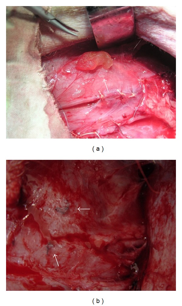
Figure 3.

(a) bFGF-CM was being implanted into the ischemic hindlimb muscle, the implanted sites were stitched as markers for samples collection. (b) At harvest, ischemic hindlimb tissue regenerated well; the stitched markers indicated the bFGF-CM implantation sites.
