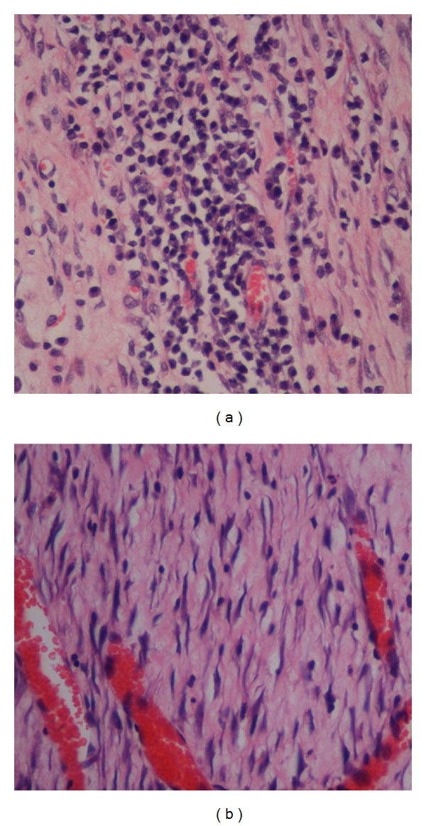
Figure 4.

High-magnification images of the histological sections of wound enclosure on day 10 after surgery in diabetic rats that are shown in Figures 3(a) and 3(d). The tissue shown in (a) harbors comparatively more inflammatory cells (mononuclear cells) than the tissue shown in (d). The tissue shown in (d) shows comparatively more collagen deposition in the wound area and increased angiogenesis relative to the tissue shown in (a) (H & E stain, magnification 40×).
