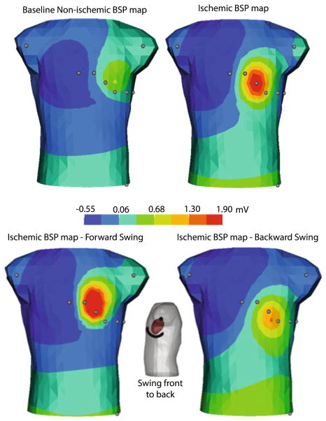
FIGURE 5.
Effect of heart position shift on ST segments during ischemic injury. The top left panel depicts the torso potentials for the baseline condition obtained from the epicardial potentials with normal blood flow. Top right panel shows the torso potentials obtained from epicardial potentials recorded during acute ischemia. The bottom row shows the torso potentials in response to the ischemic heart swinging forward and backward to reasonable physiological limits, ±17.5°.

