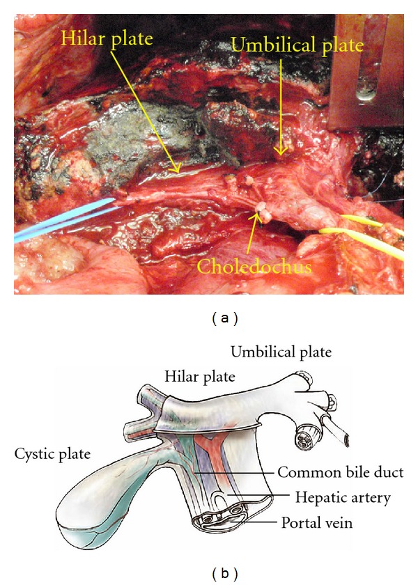
Figure 7.

The view of the hilar plate after complete eradication of the infiltrating tumor. The right portal pedicle is tapped by blue tape. The oozing blood from the liver surface is controlled by ABC. Dissection of mucinous tumor from the hilar plate after taping the right portal pedicle branches. This excision only involves surgical removal of Glisson's capsule bearing tumor and approximately 1-2 cm in depth of hepatic parenchyma.
