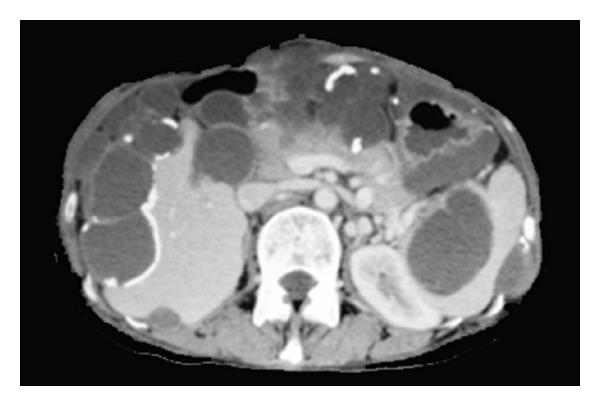
Figure 8.

Axial contrast-enhanced CT scan of the upper abdomen demonstrates multiple low attenuated cystic lesions with rim-like calcifications scalloping the liver margin, infiltrating the spleen, and compressing the bowel, pancreas, and left kidney.
